2013 Senior Thesis Abstracts
(CLASS OF 2013: If your thesis abstract is not currently included on this page and you would like it to be, please follow this link.)
The Effect of PinX1 fusion protein on telomerase activity of Xenopus laevis in vitro
Jack McTavish Craig
(Advisor: Kaplan)
Fish in the Reed Canyon have historically been the subject of great public interest and little actual research. Salmon in particular are a very charismatic species, and much of the conservation and restoration work on campus is conducted with these fish in mind. And yet, the present study is only the third thesis to conduct a thorough analysis of the Canyon's ichthyofauna, including salmonids. Annika Saltman (2008) and Laila Bryant (2009) both carried out year-long abundance monitoring regimes, and by combining their data with the new information generated here, a compelling case for multi-year trends can be made as well.
The present study followed the model of Saltman and Bryant, setting nondestructive minnow traps daily at five key sites spanning the canyon in order to gauge species abundances. Traps were set for 24 hours at five locations for 74 total trap days, totaling 8,880 total trap-hours, and almost 200 hours of field work. Importantly, this data will be directly compatible to the abundances reported in Saltman (2009) and Bryant (2008), making this the first study capable of reporting year-to-year trends. This is especially compelling in that it allows for an assessment of the Reed Canyon Enhancement Strategy, which has made significant changes to the lower Canyon since 2009 with the expressed goal of improving the habitat for fish.
It was discovered here that several fish species have increased in abundance since 2009. Red sided shiners (Richardsonius balteatus) and speckled dace (Rhinichthys osculus) both increased significantly throughout the canyon. The three spine stickleback (Gasterosteus aculeatus) and sculpin (Cottus spp.) increased significantly at Ritmanis Pond, yet stickleback actually decreased at the fish ladder. Most strikingly, juvenile Rainbow Trout (Oncorhynchus mykiss) fish were observed regularly throughout the academic year, providing the first concrete evidence that Reed Canyon is now home to salmonids.
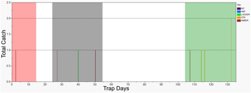
Figure 51: A timecourse of salmonid abundances at three sites.
X-axis indicates the number of days since trapping was initiated on September 27th, with shaded regions indicating times between quarters when no traps were set. Trapping sites are abbreviated as follows: RIT=Ritmanis Pond, AMP=amphitheater, LADDER=fish ladder, STR=stream, AMBER=Amber Bridge.
Proliferation and Differentiation in the Developing Zebrafish egghead Mutant Retina
Mica Emmett Peacock
(Advisor: Cerveny)
In a developing embryo, progenitor cell populations must proliferate to produce the requisite number of cells for each tissue or structure, and these cells must differentiate into adult cell-types with specialized functions. The precise control of these processes is crucial to development of a healthy adult. I investigated proliferation and differentiation in the developing zebrafish retina, which contains an adult stem cell niche called the ciliary marginal zone (CMZ). A zebrafish mutant called egghead (egh) displays a reduced and asymmetric eye phenotype at 3 days post-fertilization (dpf). The cause of this phenotype is unknown.
To determine whether changes in differentiation contribute to the egh phenotype, I performed in-situ hybridization to detect atoh7 (a marker for early retinal differentiation) in wild-type and egh embryos from 1-3 dpf. No difference in differentiation was observed between egh and wild-type individuals at any time between 1 and 3 dpf.
To investigate possible changes in proliferation contributing to the egh phenotype, I performed two types of window-labeling experiments to document the progression of progenitor cells through the cell cycle in 2.5 dpf egh and wild-type embryos. Window- labeling procedures were refined and adapted for use in the zebrafish retinal model, but fluorescent antibody detection problems prevented the acquisition of quantifiable results with which to draw conclusions regarding changes in proliferation between egh and wild-type individuals.
The direct cause of the egh phenotype is still unknown, but it appears unlikely that changes in differentiation are a significant contributor. The presence or contribution of altered cell-cycle kinetics and proliferation is unconfirmed, but remains a possibility.
Effects of the Retinoic Acid Pathway on the Behavior of Retinal Stem Cells in Zebrafish
Terra Vleeshouwer-Neumann
(Advisor: Cerveny)
Retinoic acid plays a critical role in retinal development, but the extent and mechanism of its action are far from understood. The recently identified zebrafish mutant egghead allele encodes a hypomporphic form of the protein of BMP-family member gdf6a, and can aid the investigation into retinoic acid's function. This thesis examines a few of the known phenotypes observed in the egghead-/- mutant that appear to be caused by increased retinoic acid pathway activity in the mutant retina. The reduction in gdf6a function leads to a number of retinal phenotypes, including decreased dorsal identity, reduction of the ciliary marginal zone, and ectopic "leaky" vasculature. The retinoic acid pathway was activated by exogenous retinoic acid or retinoic acid agonist Am580 application in wild type and Tg[fli1a:GFP] transgenic fish to determine its effects on the expression domain of the stem cell marker mz98, and GFP imaging provided a readout of blood vessel location in marked endothelial cells. Treatment with retinoic acid pathway agonists between 24 and 48 hours post fertilization both promoted the ectopic formation of retinal vasculature and reduced the pattern of mz98 staining. Embryos treated between 48 and 60 hours post fertilization did not show any disruption of the mz98 staining pattern. Complementary retinoic acid antagonist treatments with the retinaldehyde dehydrogenase inhibitor DEAB were performed on egghead mutants between 36 and 60 hours post fertilization, but had no effect on the staining pattern of mz98. Retinoic acid signaling interacts with the retinal stem cell niche and the developing vasculature within a specific window of time between 24 and 48 hours post fertilization to create a phenotype reminiscent of the egghead mutant, but mere inhibition of retinoic acid synthesis in egghead does not restore the wild phenotype. These results demonstrate that the retinoic acid pathway may play a role in the development of the egghead phenotype, but other defects caused by the mutation may also mediate these effects.

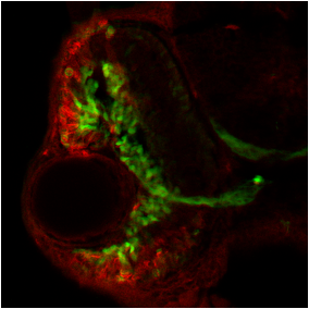
Figure 2.1 Epifluorescence microscopy of fli-GFP embryos treated from 24 to 36 hpf with RA (center), Am580 (right) or DMSO (left) as a control show potential hyper proliferation of endothelial cells in the retina, forming what appears to be excess vasculature. Eyes are oriented with dorsal toward the top of the page and ventral toward the bottom.