Under the Microscope
Winning photo from annual Developmental Biology Image Contest sheds light on development and survival.
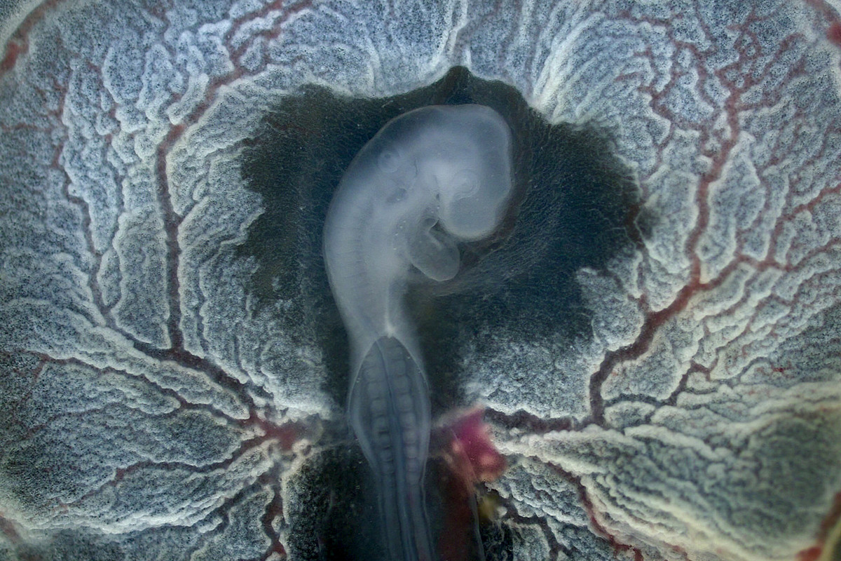
The embryo in this image belongs to a chicken strain known as an Easter Egger for the blue-and-green color of its eggs. To observe a chicken embryo, you must first crack the egg into a salt solution, remove the albumen—what we would call an egg white—and carefully peel the embryo away from the yolk.
The image was taken by mounting the embryo on the stage of a dissecting microscope, then holding a smartphone over one of the eyepieces. No longer concealed by its shell, the embryo’s heart visibly beats, pumping blood, with its head and brain oriented toward the top, and the heart tucked under its head. The membrane and blood vessels that circle the embryo provide nutrients and oxygen to support development and remove metabolic waste. In my Developmental Biology (biology 351L) lab, we study the genetic, chemical, and physical forces that influence embryo formation. This one is surprisingly resilient: as long as it is kept hydrated, covered with egg albumen, and supplied with a calcium source, it can carry on developing. The heart will continue to beat.
Tags: Academics, Object of Study