Research | Spider Development
In all spiders, early development creates a rind of cells that encloses a yolky interior. These outer cells then collect at one side of the spherical egg and the egg contracts at this side (see Movie 1). The result is formation of an embryo that looks like a ball with a flattened side. The flattened side is called the germ disc and it is these cells that will gastrulate to form the primordium of the spider’s body.
Gastrulation is a physical process critical to the normal development of all animal embryos. It converts the simple polarities of the egg into the more complex organization of the later embryo. The main body axes arise during gastrulation as a consequence of the organized movements of cells into the interior.
The canonical model states that gastrulation occurs in one continuous phase, with all cells internalizing at the blastopore at the center of the germ disc. The first cells to internalize create a small two-layered region called the primitive plate. Superficial cells continue to internalize at the center to enlarge the primitive plate, and then a group of these cells, the cumulus, detaches and migrates to the edge of the germ disc. Other primitive plate cells also migrate away from the center and are thought to form mesoderm and endoderm that will make the muscles and gut, respectively. The canonical model was derived primarily by reconstructions of fixed specimens. Movie 2 is an animation of this process. Movie 3 is a timelapse of development of Cheiracanthium mildei, a spider that fits the model.
Figure 1
Fig. 1. Canonical gastrulation compared with my laboratory’s working model of early gastrulation. Orange, primitive plate; red, cumulus; yellow, mesoderm and endoderm; blue, ectoderm. View animation.
My lab has found glaring departures from the model in three of four species by analyzing live embryos. Our model of gastrulation consists of three distinct phases, with species variation in the details: early gastrulation when the cumulus internalizes, either by itself (Movie 4) or with other primitive plate cells; mid gastrulation when the cumulus and primitive plate cells migrate away from the center (Movie 5); and late gastrulation that can occur either at the central blastopore (Movie 6) or at the periphery of the germ disc. If gastrulation occurs at the periphery, as the Oda lab (Osaka) has suggested for Parasteatoda, this would mean that in some species there are two loci of gastrulation.
After gastrulation is complete, another remarkable morphogenetic movement occurs. Inversion involves the splitting of the germ band into left and right halves. The halves move away from each other until they reach the equator (Fig 2, K–M and Movie 7). Note that the legs elongate during inversion—this can obscure the extent of inversion. Cells then migrate out of the two halves toward the dorsal midline (Fig 3) while a separate cell sheet envelops the yolk ventrally. ‘Inversion’ is an apt descriptor because cells from the outside edges of the original germ band (red in Fig 3B) meet and fuse at the dorsal midline. The spider dorsal midline, what we normally consider the central axis of an organism, originates at the very outside edges of the embryo.
Figure 2
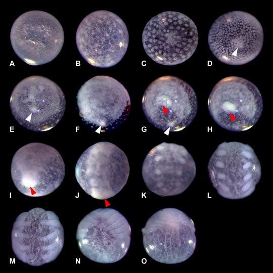
A–D, Formation of germ disc and cumulus (white arrowhead).
E–H, Migration of cumulus and formation of caudal bud (red arrowhead).
I–L, Migration of caudal bud and consolidation of germ band. Inversion begins: germ band splits into left and right halves. Appendage buds visible.
M–O, Inversion completed. Posterior body enclosed via ventral closure.
Figure 3
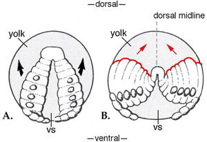
A, At the start of inversion, the germ band splits into left and right halves. The ventral sulcus (VS) enlarges as the bands migrate away from each other.
B, At the end of inversion, cells from the outside edges (red) end up at the dorsal midline.
Figure adapted from Foelix, Biology of Spiders.