
IRIS login | Reed College home Volume 93, No. 2: June 2014
Eliot Circular
Tiny Bubbles
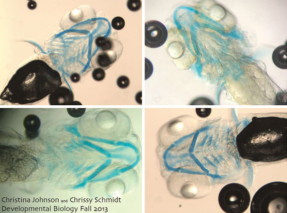
Craniofacial Cartilage, by Christina Johnson ’15 and Chrissy Schmidt ’15, shows Zebrafish embryos treated with Alcian Blue stain show craniofacial cartilage to investigate the developmental effects of valproic acid, a common anticonvulsant drug and natural developmental regulator. The dark spots in the photos are bubbles, reminiscent of the underwater bubbles.
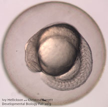
A Control, by Christina Barrett ’15 and Ivy Hellickson ’15, shows a 19-hour old zebrafish embryo that served as an untreated control for an experiment in which embryos were exposed to different amounts of gamma radiation. Although this control is normal, many irradiated embryos exhibited heart and gut abnormalities as well as decreased survival rates.
These stunning images, along with scores of others, were all captured by Reed students for independent projects in Developmental Biology (BIO 351L), taught by Prof. Kara Cerveny [biology 2012–]. Students posted the micrographs (photos taken by a camera attached to a microscope) on the department’s Dive into Development blog, which held a contest to select a winner. Nearly 300 people voted for the image they found most interesting, beautiful, and/or provocative. The two images showcased here each received 82 votes to tie for first place.
Editor's Note
Features
- Distant Vision
- Looking Back at Freedom Summer
- Life Beyond Reed
- Into the Unknown
- Stroke of Genius
- The Long Run
- Behind the Curve
- Cliff Hanger
- Keeping the Flame
- Poet || Artist
- Why I Don’t Give to Reed
Reediana
- Barbara Ehrenreich ’63: Living with a Wild God
- Aaron Glass ’94: Return to the Land of the Head Hunters
- Lee Oser ’86: The Oracles Fell Silent
- Prof. Douglas Fix: Notes on Travel in Formosa
- Reediana Briefs
Eliot Circular
- Reed’s First Building Slated for Demolition
- Professors Corner
- STEM Femmes Arise
- New Faces
- Scholarship named for Abby Garcia ’10
- English Prof. Turns 100
- Tiny Bubbles
- Final Stanza
LATEST COMMENTS
steve-jobs-1976 I knew Steve Jobs when he was on the second floor of Quincy. (Fall...
Utnapishtim - 2 weeks ago
Prof. Mason Drukman [political science 1964–70] This is gold, pure gold. God bless, Prof. Drukman.
puredog - 1 month ago
virginia-davis-1965 Such a good friend & compatriot in the day of Satyricon...
czarchasm - 4 months ago
John Peara Baba 1990 John died of a broken heart from losing his mom and then his...
kodachrome - 7 months ago
Carol Sawyer 1962 Who wrote this obit? I'm writing something about Carol Sawyer...
MsLaurie Pepper - 8 months ago
William W. Wissman MAT 1969 ...and THREE sisters. Sabra, the oldest, Mary, the middle, and...
riclf - 10 months ago