Renn Lab Thesis Project
Cadence True
Social Regulation of Neuropeptides in Astatotilapia burtoni
A variety of hormones and neuropeptides have been shown to
regulate reproductive behaviors but the mechanism for this regulation
is still an important question requiring further research. In the African
cichlid fish Astatotilapia burtoni males can be of a territorial or non-territorial
phenotype. Territorial (T) males defend a territory and spawn with females
while non-territorial (NT) males are reproductively repressed in behavior
and physiology. This example of alternative male reproductive tactics
has been well studied on the hormonal and molecular level and represents
an ideal model for investigating the mechanisms that control reproduction,
specifically courtship behaviors as well as physiological capacity. Another
reason this teleost species represents a good opportunity to study neural
control of behavior is because the new technology of microarrays has
been applied to this species to screen for gene expression differences
between T and NT males. Among some of these genes are many neurotransmitter
proteins, including GABA and kainate receptors as well as the neuropeptides
gonadotropin-releasing hormone (GnRH) and arginine vasotocin (AVT). Microarray
results only indicate whole brain increases in GnRH and AVT mRNA within
T males and the localization of these peptide differences is important
for understanding the significance and regulatory effects of these molecules.
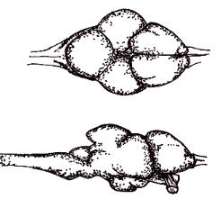
Here immunocytochemistry was used to localize both GnRH and AVT neurons
in A. burtoni. 
Territorial males showed larger numbers of AVT cells than NT males in the preoptic area. Importantly, it was discovered that a subpopulation of AVT neurons also contained GnRH. This is the first example of colocalization of these two neuropeptides and indicates a possible interaction for these molecules in regulating reproductive behavior and physiology.
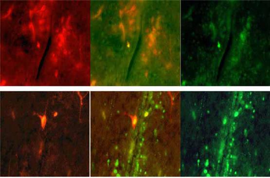
Figure 2. Colocalizations of Arginine Vasotocin and Gonadotropin-Releasing Hormone within POA cells of A. burtoni. Arginine vasotocin (AVT) was identified using immunocytochemistry with a fluorescent secondary antibody that fluoresced red (left panels), while gonadotropin-releasing hormone (GnRH) had a secondary antibody that fluoresced green (right panels). Using Photoshop it is possible to overlap pictures taken from each channel to produce an image of fluorescing cells from both channels (middle panel). Shown here are two instances in which some cells appear double labeled (yellow) while other cells appear to only be positive for AVT.
Click HERE to see other Reed Biology Theses.