Sound Wall
Work in Exhibition
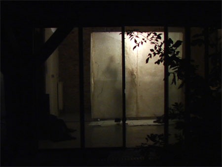
Sound Wall, 2008
Hand-made abaca paper, speaker embedded in paper, MP3 players, aluminum frame 24’ x 8’.
The sound of cells emanates from small speakers embedded in the 24’ wall of handmade paper that curves around the gallery. The sounds are from cells, both healthy and ill, dividing and dying. Bio-physicist Andrew Pelling at UCLA made the recording using an Atomic Force Microscope. To render such minute units audible, the nanoscale vibrations of cells are amplified and brought into the range of human hearing. Pelling found that cells with cancer or other diseases give off low and strained-sounding frequencies, while healthy cells produce a more even-toned sound.
Artist Statement
The cell, the smallest unit in a living organism, houses genetic code. Both an object and a vessel, it is a site of activity that is never static or complete. The term cell comes from the Latin cella, meaning “small room”. Historically the architectural cell is both a space for reverent solitude and meditation, or for captivity and confinement. The biological association was given by the British scholar Robert Hooke in 1662. The first to use microscopic magnification to study and render cellular structure, Hooke noted that cork cells resembled the small rooms monks lived in. His etchings of magnified organic forms published in Micrographia in 1665, revolutionized the way in which biological organisms were understood. Optics and magnification have continued to give vision to the previously unseen and have changed our understanding of interior systems.
Most recently, nanotechnology, particularly the AFM (atomic force microscope) pioneered by James Gimzewski, Professor of Chemistry at UCLA and Andrew Pelling, Associate Professor of Biophysics at the University of Ottawa, have enabled researchers to touch and to listen to cells. Like the blind reading braille or a finger] touching a pulse, the needle of the AMF can touch and feel the vibrations of cells less than half the diameter of a human hair. The new area of study is called sonocytology, which uses the atomic force microscope (AFM) to feel a cell in the same way a needle was used to feel the pattern of vibrations pressed into vinyl records. Gimzewski and Pelling have found that cells with cancer or other diseases emit a very low and strained frequency, while healthy cells produce a more pleasant sound. Sonocytology has proven to be a noninvasive way to detect disease. In 2007, Gimzewski used nanotechnology to demonstrate that metastasized cancer cells are softer than healthy cells. The study represented one of the first times researchers have been able to take living cells from human cancer patients and use nanotechnology to determine which were cancerous through touch.
Through touching and hearing rather than only seeing, an entirely new way of studying a form—that of the multiple sensory knowledge—has been born. This ability to discern cellular soundings has ultimately revolutionized the understanding of cellular gestation, cell division, and the relationships within the interior architecture of our body.
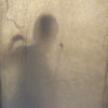
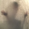
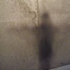
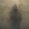
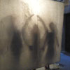
When I learned of this phenomenal research done at UCLA, I began to consider touch and sound as vital elements in creating a work about cells. Over the past two years I have spent time listing to and learning about the sounds of cells, and the pace and visual nuance of cell division. As a result, I created the work Sound Wall as a physical, multi-sensory experience. Using the main space of the Western Gallery as a vessel, I designed three cellular spaces--one, 7’ x 14’ x 8’, a second 6’ x 12’ x 8’ and a third 8’ x 4’ x 8’--for the viewers to move in and out of. These minimal cubic forms are layered, one in front of the other, so that one can see the physical ratio of the three spaces simultaneously. The spaces, made from handmade abaca paper are illuminated from the interior to create a lantern-like effect. When inside the space the viewers’ bodies project distorted shadows as they feel and listen to the sounds of cells dividing and emanating from tiny speakers embedded in the walls of paper.
The sounds one is hearing and feeling are five tracks of healthy to damaged cells, dividing and dying, layered with my own recording of a monastery cell, and family conversations. I was permitted the copyright for these five recordings by Andrew Pelling. The sound recordings used for this work range from rhythmic to strained. The pitch of each is so distinct that even the untrained ear can sense the sound of health and distress as one moves through and touches the thin handmade paper walls.
I chose to make this work with handmade abaca paper, because like the cellular walls, it is both translucent and penetrable. Both paper and cells are vehicles that hold and convey fundamental information on the interior life of others. Abaca paper, made from palm leaf since the 14th century, has been traditionally used for letter writing, drafting, typewriters, and rope-making—all materials used for communication and uniting. It has remarkable tensile strength, which made it possible to cast 4’ x 8’ panels, sew them into large wall sections and frame them into aluminum structures.
To make 4’ x 8’ sheets of paper I poured thoroughly beaten pulp on to a 4’ x 8’ cedar silkscreen frame system. A very thin sheet of plastic lined the frame so that once the pulp was poured in, the plastic could be removed, and pulp could flow over the silk evenly. The processes of pouring and the bonding and drying of pulped plant fiber is evident in each sheet that makes up the walls of this work. As a result, the paper looks very much like the cellular wall one sees through the microscope.
Special Thanks
Andrew E. Pelling, Assistant Professor of Biophysics, Department of Physics, University of Ottawa, for his sound recording of the cells as used in Dark Side of the Cell by Anne Niemetz and Andrew Pelling 2004. Dominic Redferm, Senior Lecturer of Media Arts at RMIT, for shooting and editing the video. Siobhan Murphy, Choreographer, University of Victoria, and Greg Miller, Professor of English and Poet, Millsap College, for their choreography, singing and performance. This project was funded by the Mellon Foundation for Faculty Research and Development, the Stillman-Drake Fund, Reed College, CAMAC, and the Tenot Foundation
