
IRIS login | Reed College home Volume 96, No. 1: March 2017
Object of Study
Eye of the Beholder
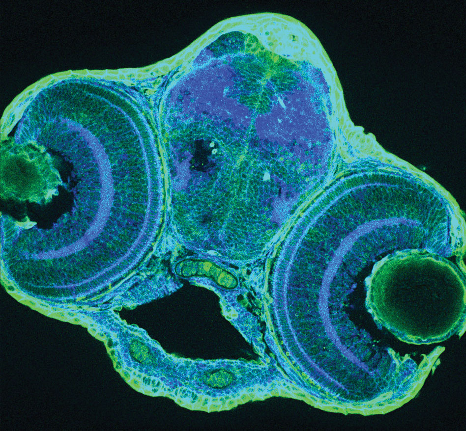
Students in Prof. Kara Cerveny’s Developmental Biology class (Bio 351) study how neural stem and progenitor cells transition from proliferation to differentiation—that is, from being “generic” cells to being specialized cells, like blood cells, bone cells, and brain cells.
Students investigate this crucial event in the visual system of zebrafish, using cellular, genetic, and embryological manipulations to probe how progenitor cells regulate their behaviors by integrating extrinsic signals with cell-intrinsic cues. In the lab, students cut thin slices of zebrafish embryos and stain them with antibodies to examine cellular architecture.
Here we see a transverse cross-section from the head of a three-day-old zebrafish embryo stained with antibodies for beta-catenin (blue) and green fluorescent protein (um, green). The central open space is the mouth and the tissue directly above it is the forebrain. On either side of the forebrain sit the two eyes, each comprised of a spherical lens and a trilaminar multicellular retina. The blue blobs in the forebrain highlight regions where many neurons connect with each other. This image was captured on a laser-scanning Nikon A1+ confocal with a 25X 1.2 NA water dipping lens by Avery Van Duzer ’18 and Evan Welch ’17.
Editor's Note
Features
- Breaking the Frame
- Codebreaker
- The Dynamics of Compassion
- Taking a Fresh Look at Hum 110
- Chasing Coyote
- Eye of the Beholder
LATEST COMMENTS
steve-jobs-1976 I knew Steve Jobs when he was on the second floor of Quincy. (Fall...
Utnapishtim - 2 weeks ago
Prof. Mason Drukman [political science 1964–70] This is gold, pure gold. God bless, Prof. Drukman.
puredog - 1 month ago
virginia-davis-1965 Such a good friend & compatriot in the day of Satyricon...
czarchasm - 4 months ago
John Peara Baba 1990 John died of a broken heart from losing his mom and then his...
kodachrome - 7 months ago
Carol Sawyer 1962 Who wrote this obit? I'm writing something about Carol Sawyer...
MsLaurie Pepper - 8 months ago
William W. Wissman MAT 1969 ...and THREE sisters. Sabra, the oldest, Mary, the middle, and...
riclf - 10 months ago