The Jewel Wasp
Biology 342 Fall 2010
Briana Patton and Lisa Schomaker
Envenomation
World's Smallest Surgeon and Anesthesiologist
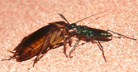 The wasp achieves behavioral control of the cockroach by selectively tampering with its neural circuitry. With surgical precision “akin to the most advanced sterotactic delivery of drugs,” the wasp injects a venom cocktail into the cerebral ganglia of the cockroach, which over the course of an hour or so, renders the cockroach a 'zombie,' that does the wasp's bidding. Prior to injecting venom into the roach's brain, the wasp anesthetizes the cockroach by stinging it in the thorax, producing a temporary paralysis and enabling the wasp to accurately place the second sting. During paralysis, the venom acts directly at the neuromuscular junctions of the thoracic ganglion to prevent movement. (Figure 4. Ampulex compressa leading a subdued cockroach toward a nesting burrow . Photo source 2)
Neural Hijacking
During the temporary paralysis, the wasp then stings the head ganglia, injecting venom directly into the brain. The venom delivery site can be visualized by injecting wasps with radiolabeled amino acids (which are incorporated into the venom) and tracking its localization in stung cockroaches. This approach revealed that the injected venom is concentrated in the head ganglia, clusters of neuronal tissue that are divided into the brain (also called the supraesophageal ganglion, or SupEG) and the subesophageal ganglion (SEG, refer to figure 4). The wasp targets certain structures within the head ganglia, as evidenced by particularly high concentrations of radiolabeled venom found in the central aspect of the brain around the mushroom bodies, and in the midline of the SEG. These regions control important aspects of the cockroach's higher functioning.
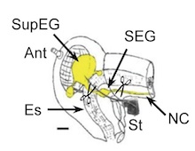
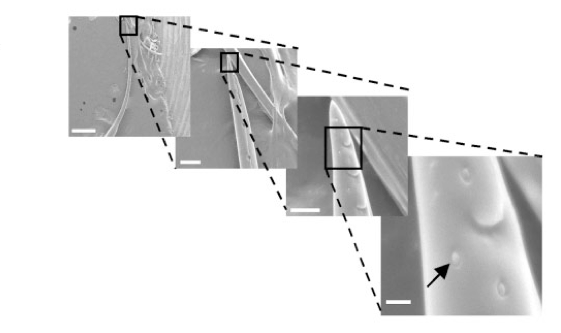
Figure 4. (left) Neuroanatomy of a Periplaneta americana (right) electron microscopy of sensilla on the stinger of a jewel wasp (Gal R. et al. 2005)
The degree of specificity with which the wasp stings may be aided by specialized sensors located on its stinger. A 2005 study by fiGal et al., which compared “sting duration for wasps stinging sham operated roaches to those of Figure 2. Cockroach neuroanatomy brainless roaches, in which the brain was removed prior to the sting” showed that wasps can discriminate between neural and non neural tissue. Wasps that stung brainless cockroaches did so for 15 fold longer, and deposited little venom in comparison to wasps stinging sham operated roaches (Gal R. et al. 2005). Electron scanning microscopy of the wasp's stinger found structures resembling sensilla, insect sensory organs, on the most distal portion of the stinger (refer to Figure 4.), though it is unknown what cues the sensilla would be receiving in order to detect neural tissue.