Renn Lab Thesis Project
2012 Reed Graduate
MORGAN MOVIUS
Neuropeptides and sex-biased behavior: Arginine Vasotocin and Isotocin in Julidochromis
In many vertebrate species behavioral phenotypes are highly sex-biased, and one sex typically displays territorial and aggressive behaviors while the other sex exhibits more submissive and parental behaviors. Julidochromis marlieri and Julidochromis transcriptus are two closely related cichlid fish species that have evolved opposite patterns of sex-biased behaviors since diverging from their common ancestor. J. transcriptus exhibits conventional sex-biased behavior in which the male is highly territorial and aggressive and the female is submissive. J. marlieri exhibits a pattern of sex-biased behavior in which the female is more aggressive and the male is submissive.
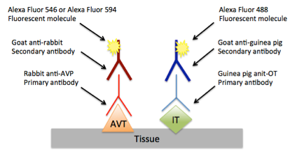
Previous gene expression research has shown that aggressive J. transcriptus males and aggressive J. marlieri females both have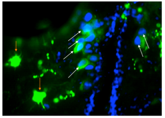 elevated levels arginine vasotocin (AVT) and isotocin (IT) mRNA when compared with their subordinate conspecifics, suggesting that these peptides may contribute to the formation of aggressive behavioral phenotypes in both species.
Figure 1: Cell number was determined by the presence of both green fluorescence from the Alexa488 secondary antibody and blue DAPI nuclei. Red arrows indicate fluorescent bodies not considered cells due to the lack of nuclei. White arrows indicate fluorescent bodies counted as cells. Image taken by author.
The current study aims to characterize the expression of AVT and IT proteins on a cellular level in the preoptic area of the central nervous system.
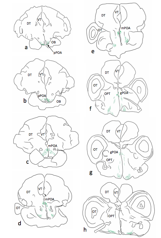
Figure 2: Representative series of line drawings from a J. marlieri female illustrating the distribution of IT-immunoreactive cells and terminals within the brain. IT-immunoreactive cells are represented as green circles, IT terminals are represented as solid green lines. Abbreviations: VT: ventral telencephalon; DT: dorsal telencephalon; OT: optic tectum; OPT; optic tract; pPOA: parvocelular preoptic area; mPOA: magnocellular preoptic area; gPOA: giantocellular preoptic area. Images created by author.
AVT and IT expressing cells will compared based on both size and number in an effort to explain the mRNA results on a cellular level. Cell size and cell number were measured for each individual animal and compared by species, sex, and behavioral phenotype. While cell size was not significantly different between species (Figure 3A), sex, or behavioral phenotype, J. marlieri females had more isotocin producing cells than any other experimental group, including J. transcriptus males (Figure 3B).
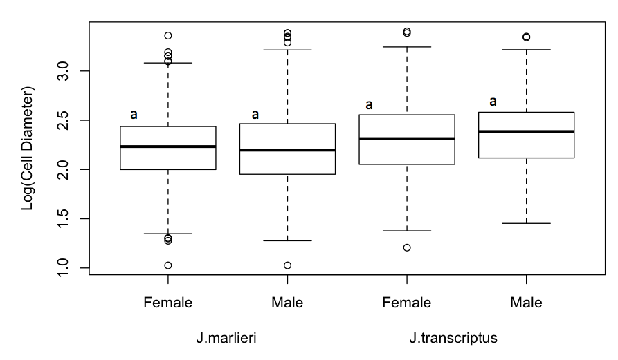
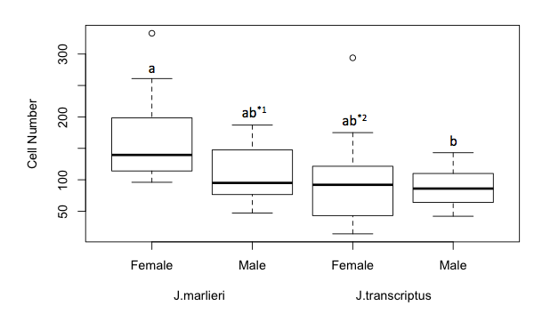
Figure 3: A) Cell diameter analyzed by group. Boxplots annotated with different letters indicate groups that are statistically different (p≤0.05). No comparison between groups was statistically significant. B) Cell number comparisons between groups. Boxplots annotated with different letters indicate groups that are statistically different (p≤0.05). For b, p=0.029 for the difference between J. transcriptus males and J. marlieri females; ab*2, p=0.052 for the difference between J. transcriptus females and J. marlieri females; ab*1, p=0.12 for the difference between J. marlieri males and J. marlieri females.
This research suggests that J. marlieri females increase isotocin expression by increasing the number of isotocin producing cells in the brain. However, it is unclear how J. transcriptus males increase isotocin expression, since these animals had neither more nor larger isotocin producing cells. The data from the current research suggest that J. transcriptus males and J. marlieri females have evolved different cellular mechanisms to increase the expression of isotocin. Therefore, it appears that similar behavioral phenotypes may be achieved through divergent mechanisms in these fishes.