South American Gynotiforms: Conductance as Communication
Biology 342 Fall 2011
Moriah Gottman and Misha Naiman
There are a wide array of gymnotiforms which develop electric organs in different ways. The paper "Ontogeny and evolution of electric organs in Gymnotiform fish" by Kirshbaum and Schwassmann compares the development of electric organs in eight different species including Eigenmannia virescens, Sternopygus macrurus, and Electrophorus electricus. These eight species can be separated into two broadly defined developmental groups, with one group developing type A electrocytes and the other developing type B electrocytes.
Species that develop type A electrocytes include the Eigenmannia vierscens and Sternopyguy marcrurus. These species send out wave type electric discharge, which is defined as a continuous pulse with the fish capable of changing the frequency of the pulse. Kirschbaum and Schwassmann seem to think wave type discharge is the plesiomorphic condition (or a condition shared with the ancestral clade) because of the electrocytes' physiological similarity to the muscles fibers surrounding them, and the way electrocytes develop densly within the hypaxial muscles. The larvae in this group first start developing electrocytes as early as 7 days after hatching. They begin their developmental cycle by expressing type A electroblasts directly beneath the hypaxial muscle, which disperse within the muscles as the electroblasts mature. As fry grow bigger, the electrocytes shift from the rostral part of the fish to a more caudal location or disappear for good. The shift of early electrocytes from rostral to a more caudal location is something that occured in the evolution of the African mormyrid, a completely different phylum of electric fish.
Other species including the Electrophorus electricus and Gymnotiform carapo, develope type B electrocytes, which tend to send out pulse type electric discharge. Pulse type electric discharge can be defined as a infrequent pulse which is sent out of the electric organ of the fish to the surrounding environment at a steady frequency. Larvae start showing showing signs of electrocyte B development 7 days after hatching, with electric organs becoming more sophisticated with maturation. Species with type B electrocytes tend to develope preelectrocytes from 4-9 days after hatchings, which are capable of emmitting very weak monophasic charges. These charges become stronger and develop a more specified pattern as the larvae mature, eventually becoming the main form of fish-fish communication as well as fish-environment communication. In the two gymnotiform species the electrocytes extended dorsally into the hypaxial muscle, while the pre-electrocytes of other species like those of the Rhamphichthys sp. develop directly into adult electric organs. The Electrophorus electricus, as it develops, forms two different electric organs that are called Sack's organ and Hunter's organ, which other electric fish don't have. This phenomenon could be due to the fact that Electrophorus electricus has the strongest electric discharge of any type of electric fish.
All species of gymnotiforms produce a monophasic discharge during their larval stage, and as the fish grow older the electric discharge produced becomes more complex as it fulfills the roles of communication, echolocation, and mating signals in adult gymnotiforms.
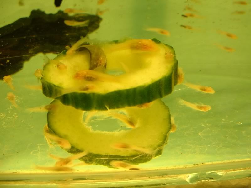
Figure 4. Gymnotiform fry, newly hatched, in a standard lab-issue tank.
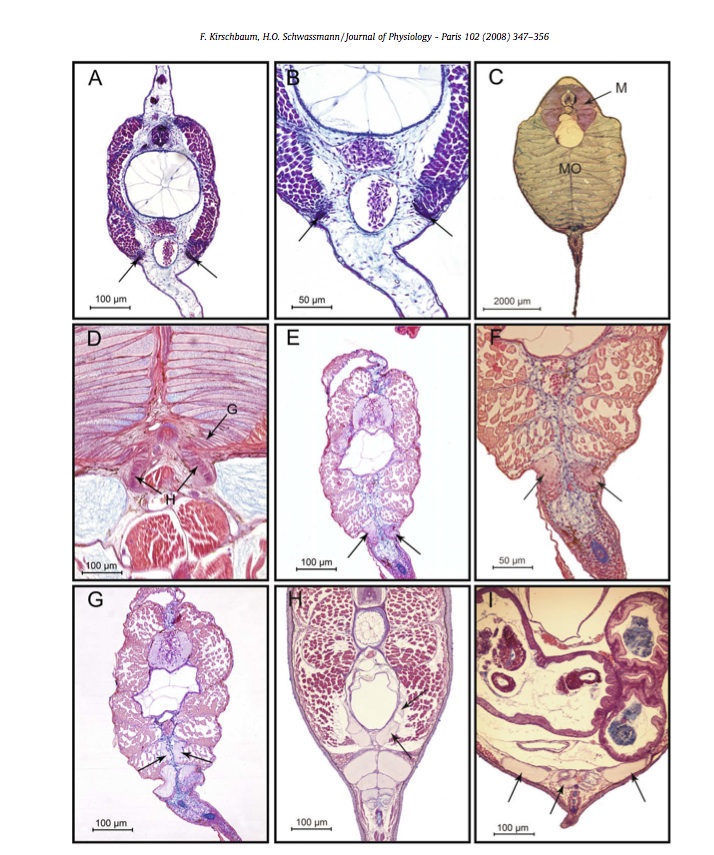
Figure 5. Micrographs of disected electric organs of Electrophorous electricus (A-D), Gymnotus aff. carapo (E-G), and G. carapo (H-I). Images A-D display the electric organs of four hatchlings- the A and B hatchlings are 10mm long each and the image shows pre-electrocytes ventral to the hypaxial muscle (arrows), C is a 70 mm long adolescent whose electric organ is mostly filled with the gap below the hypaxial muscle, D is a 140mm long juvenille whose electrical organ demonstrates a splitting off of additional electrocytes ventral to the germanitive region that lead to the development of the Hunter's organ ventral to the hypaxial muscle(H,G), E-G are juvenilles also 140mm long whose pre-electrocytes are below the hypaxial muscle (arrows), H's juvenille electrocytes extend completely into the hypaxial muscle, and I is a young adult whose electrocytes are located ventral to the body cavity and rostral on the body.